
A complete blood count with differential is recommended in patients with a history and physical examination suggestive of infection or malignancy however, good evidence to support the value of routine complete blood count is lacking. Results of a complete blood count with differential may be abnormal with infectious lymphadenitis. Ordering routine studies in a shotgun style approach is rarely indicated and seldom can reliably rule in or out a specific disease ( Table 3). Laboratory studies may be indicated if there is concern about a systemic disease or to confirm a diagnosis suspected from the history and physical examination. The primary care physician ultimately must determine whether further invasive workup or treatment is necessary, or if watchful waiting is appropriate. If malignancy is suspected (accompanying type B symptoms hard, firm, or rubbery consistency fixed mass supraclavicular mass lymph node larger than 2 cm in diameter persistent enlargement for more than two weeks no decrease in size after four to six weeks absence of inflammation ulceration failure to respond to antibiotic therapy or a thyroid mass), the patient should be referred to a head and neck surgeon for urgent evaluation and possible biopsy. Lack of response to initial antibiotics should prompt consideration of intravenous antibiotic therapy, referral for possible incision and drainage, or further workup.

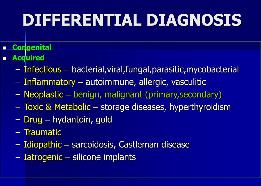
Antibiotic therapy for suspected bacterial lymphadenitis should target Staphylococcus aureus and group A streptococcus. Congenital neck masses are excised to prevent potential growth and secondary infection of the lesion. Computed tomography with intravenous contrast media is recommended for evaluating a malignancy or a suspected retropharyngeal or deep neck abscess. Ultrasonography is the preferred imaging study for a developmental or palpable mass. Workup for a neck mass may include a complete blood count purified protein derivative test for tuberculosis and measurement of titers for Epstein-Barr virus, cat-scratch disease, cytomegalovirus, human immunodeficiency virus, and toxoplasmosis if the history raises suspicion for any of these conditions. Although rare in children, malignant lesions occurring in the neck include lymphoma, rhabdomyosarcoma, thyroid carcinoma, and metastatic nasopharyngeal carcinoma. Common benign neoplastic lesions include pilomatrixomas, lipomas, fibromas, neurofibromas, and salivary gland tumors. Inflammatory neck masses can be the result of reactive lymphadenopathy, infectious lymphadenitis (viral, staphylococcal, and mycobacterial infections cat-scratch disease), or Kawasaki disease. Common congenital developmental masses in the neck include thyroglossal duct cysts, branchial cleft cysts, dermoid cysts, vascular malformations, and hemangiomas. Neck masses in children usually fall into one of three categories: developmental, inflammatory/reactive, or neoplastic.


 0 kommentar(er)
